THE BASIC HUMAN COLOR VISION SYSTEM
The visible Spectrum
The visible spectrum is the portion of the electromagnetic spectrum with wavelengths between 380 nm and 760 nm. These wavelengths (and frequencies) are sensed as the following colors (wavelengths are the centers of the named colors):
- 400 nm (or 750 THz) - violet
- 460 nm (or 650 THz) - blue
- 490 nm (or 610 THz) - cyan
- 520 nm (or 580 THz) - green
- 570 nm (or 530 THz) - yellow
- 590 nm (or 510 THz) - orange
- 670 nm (or 450 THz) - red
Four kinds of sensors:
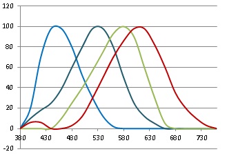
Use the chart of light sensitivity curves at right.
-
Rods
Rods are primarily used at night, because they are much more sensitive than the cones. But they also provide motion and edge detection in brighter light. The rhodopsin (visual purple) in the rods is sensitive to a range of wavelengths between 380 nm and 590 nm, peaking at 510 nm. This covers the colors violet, blue, cyan, green, yellow, and orange. But rods are encoded as white, not as any specific color, because they serve for night vision.
-
Blue-Sensitive Cones
The cyanopsin in the blue-sensitive cones is sensitive to a range of wavelengths between 380 nm and 550 nm, peaking at 450 nm. This includes violet, blue, and cyan light.
-
Green-Sensitive Cones
The chloropsin in the green-sensitive cones is sensitive to a range of wavelengths between 430 nm and 670 nm, peaking at 550 nm. This includes cyan, green, yellow, and orange light.
-
Red-Sensitive Cones
The erythropsin in the red-sensitive cones is sensitive to two ranges of wavelengths. The major range is between 500 nm and 760 nm, peaking at 600 nm. This includes green, yellow, orange, and red light. The minor range is between 380 nm and 450 nm, peaking at 420 nm. This includes violet and some blue. The minor range is what makes the hues appear to form a circle instead of a straight line.
Seeing other colors:
- Violet activates the blue cone, and partially activates the red cone.
- Blue activates the blue cone.
- Cyan activates the blue cone and the green cone.
- Green activates the green cone, and slightly activates the red and blue cones.
- Yellow activates the green cone and the red cone.
- Orange activates the red cone, and slightly activates the green cone.
- Red activates the red cone.
- Magenta activates the red cone and the blue cone.
- White activates the red cone, the green cone, and the blue cone.
Colors not listed here are seen due to varying strengths of light activating the red, green, and blue cones. A few examples follow:
- Black does not activate any of the cones.
- Brown partially activates the red cone, and the green cone a little less.
- Brick Red partially activates red cone.
- Pink activates the red cone, and partially activates the green and blue cones.
- Flesh fully activates red, activates green a little less, and blue a little less than that.
- Amber activates the red cone, and green a little less.
- Ochre partially activates the red cone, and green a little less.
- Olive partially activates the green cone, and red a little less.
- Gray partially activates the red cone, the green cone, and the blue cone.
What happens at night - Rod vision:
- Violet activates the rods partially
- Blue activates the rods almost fully.
- Cyan activates the rods fully.
- Green activates the rods fully.
- Yellow activates the rods almost fully.
- Orange activates the rods partially.
- Red does not activate the rods.
- Magenta activates the rods partially.
- White activates the rods fully.
After the cones have received the colors, the color information is encoded in the bipolar and ganglion cells in the retina before it is passed on to the brain. Three different encodings are used:
- The primary encoding is luminance (brightness). It is the sum of the signals coming from the red cones, the green cones, the blue cones, and the rods. These provide the fine detail of the image in black and white.
- A second form of encoding is used to separate blue and yellow (yellow being the sum of red and green). The signal changes one way if blue is stronger than yellow, and changes in the opposite way if yellow is stronger than blue. This is blue-yellow.
- The third form of encoding is used to separate red and green. The signal changes one way if green is stronger than red, and changes the other way if red is stronger than green. This is green-red.
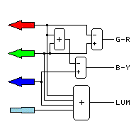
The outputs of the eye's color encoding matrix are shown to the right:
- The blue-yellow axis is horizontal.
- The green-red axis is vertical.
The second and third kind of encoding do not have quite the fine resolution the luminance encoding has. Black and white vision has finer detail than color vision.
In the fovea, where central vision occurs, each luminance ganglion cell receives signal from only one cone cell of each color. There are no rod cells in the fovea, so it is night blind.
It is interesting that in the very center of the fovea, there are also no blue-sensitive cells and few green-sensitive cells. This area gets its color information from cells surrounding it.
An averaging mechanism is used in the brain. It creates a norm for an area around the item being looked at. Three norms are created, one for light intensity, one for blue-yellow difference, and one for red-green difference. The colors of objects are compared to the norms, cancelling the color of the light out of the color of the item where possible (see below).
This color matrix is responsible for the psychological primaries (see below). They are found at the ends of the center column and the center row (at right).
COLOR MATRIX OUTPUTS
. yel − mid
+ blu .
grn
grn
+
+
mid
mid
−
−
red
red
. yel − mid
+ blu .
| Psychological Primaries | |||
|---|---|---|---|
| cerise | yellow | aqua | blue |
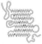
Note that the genes for the cone cells have two segments.
- One segment encodes a protein making an alternating grid with the proper spacing to be
affected by certain colors of light. The rods and the different kinds of cone cells have
different grid spacings, so they are sensitive to different colors.
A rough diagram of the grid is shown at right. Notice the equally spaced rows of alpha-helix configured amino acids in the molecule. The spacings line up with the wavelength of the light of the color the molecule is sensitive too. The light effectively rattles the entire molecule if it is the right color.
- The other segment of the gene tells the cone cell which bipolar cell nerve endings to connect to, so the color signals are properly encoded to be sent to the brain.
It is interesting that the light has to pass through the ganglion and bipolar cells to get to the rods and cones. The retina seems to be made backward. But this might protect the light-sensitive cells from damage.
HOW HUMAN VISION REACTS TO DIFFERENT LIGHT SOURCES
The human visual system reacts in three different ways to three different kinds of light.
Here the effects of the averaging system (mentioned earlier) can be seen. If the light source allows it, the human visual system cancels out the color of the light source.
Notice how, although the eye compensates for the color of the light source (if it can), the camera does not. The color of the light is compensated for by the photographer, by selecting an indoor or outdoor film, or through darkroom or electronic techniques.
- Smooth lighting curves:
Human color vision has the ability to automatically adjust itself to lighting of different color temperatures, provided the lighting has a smooth (black-body) nature. Examples of black body light include sunlight, incandescent lamps, candles, campfires, and changed sunlight, due to sunset, an overcast sky, or the northern sky being the primary source of light.
The brain compares the ratio of red to blue for each object to the ratio of red to blue in the averaged light. The viewer is unaware of any color shift due to the difference in the light. But since cameras do not make this correction, the viewer is surprised when the colors look so strange on the prints.
Smooth Lighting Curve Examples:
These look right to the eye, not cameras:
Blue White Green Yellow Red
Blue White Green Yellow Red
Blue White Green Yellow Red
- Choppy lighting curve:
If the light source contains bright lines or gaps in the spectrum, the human vision system can not correct the colors the way it can for changes in color temperature. Examples of this kind of light include mercury vapor light, some kinds of fluorescent light, and high pressure sodium vapor light.
Notice that some colors are not seen as the correct color. Red is entirely absent from mercury vapor light, and both green and blue are absent from high pressure sodium vapor light. The colors change to show what light the object can reflect. In this case, the camera may have the same troubles they have in the previous case.
Choppy Lighting Curve Examples:
These do not look right:
Blue White Green Yellow Red
Blue White Green Yellow Red
- Low level light, or light that is nearly all one color:
When the light level is low, only one light sensor is activated. Everything looks like shades of gray. Red objects look darker than green or blue, because the rods are totally insensitive to red. Cameras produce totally unpredictable results under low light. Often the photos are in a single primary color.
In the case of low pressure sodium, all of the light is yellow, so it activates both the red and green cone. But everything illuminated by this light returns the same wavelength of yellow, so there is no difference in color. The only difference between the light reflected by different objects is brightness. Objects that are not yellow, and are not tints of other colors, look black. In this case, the camera produces the same result the eye does.
Other monochromatic lights produce similar results, with a different color being the only real difference.
Single Color Curve Examples:
Monochromatic or low level light:
Blue White Green Yellow Red
Blue White Green Yellow Red
HOW HUMAN COLOR VISION DICTATES PRIMARY COLORS
Light Primaries
The key to the primary colors of light is the set of response curves of the cones. Note the
place where each cone has the least overlap with other cones.

670 nm red
530 nm green
440 nm blue
These are the primary colors of light. They are defined by the response curves themselves. But note that the peak of the response curve does not define the primary color. Each wavelength where a cone is acting mostly alone determines a primary color.
Human Cone
Response Curves
Wrong Primaries
 Some die-hards stick to the old set of pigment primary colors:
670 nm red
578 nm yellow
440 nm blue
But they work only if they are leaky, and they produce a much smaller gamut of colors. These colors are impossible to make in this system:
Those who adhere to this obsolete set of primary colors do so because "the masters" used them. The masters used what they could get at the time. Magenta and cyan were not available in permanent pigment form then.
This system works better in oil paint than it does in other media, because:
- The best blue becomes cyan at low concentrations
- The best red becomes magenta at low concentrations
- Some oil paints mix as light mixes, instead of as pigment mixes
But it does not work in other media without using leaky pigments. Equal amounts of all three leaky pigments make brown instead of black, showing the defects in the system.
The biggest problem is that art teachers in schools are still teaching the old system - even as they watch it fail when they try to teach it with crayons or water colors. And Crayola™ sells products designed for the old system, but not for the new system.
Light Secondaries
To find the secondary colors, find the colors that are produced by equal mixtures of each pair of primaries:
Cyan is produced by an equal mixture of blue
and green light.
Magenta is an equal mixture of blue and
red light.
Yellow is produced by an equal mixture of
green and red light.
Equally mixing red, green, and blue light produces a sensation equal to white light.
Pigment Primaries
 The key to finding the primary colors of pigment is to find 3 pigments, each one absorbing only
one primary color of light.
The key to finding the primary colors of pigment is to find 3 pigments, each one absorbing only
one primary color of light.
Cyan removes red light, but not blue or green
light.
Magenta removes green light, but not blue
or red light.
Yellow removes blue light, but not green
or red light.
Equally mixing cyan, magenta, and yellow pigments produces black.
Thus, the secondary colors of light are the colors that make a very good set of pigment primaries. And the secondary colors of pigment are the light primaries.
Note that most kinds of defective color vision need different sets of primary colors. Dichromatic vision has only two primaries.
Pigment Secondaries
The pigment secondaries are the light primaries.
Psychological Primaries
A third set of primary colors comes from the color encoding matrix in the eye (above):
cerise
yellow
aqua
blue
These do not work for mixing of either pigments or lights. Their use is confined to the brain responses to color stimuli.
Mistakes
The following colors are often mistaken for each other, not because of problems with human vision, but because of problems in human language. Many people do not know what "magenta" and "cyan" are, or that they are distinct colors:
- Set 1 is often mistakenly grouped as "red" by those who do not understand color
theory:
Red light is primary, containing only red.
Magenta is an equal mixture of blue and red light. - Set 2 is often mistakenly grouped as "pink" by those who do not understand color
theory:
Pink is a mixture of strong red light with weaker light of all other colors.
Magenta is an equal mixture of blue and red light. - Set 3 is often mistakenly grouped as "blue" by those who do not understand color
theory:
Blue light is primary, containing only blue.
Cyan is an equal mixture of blue and green light.
These mistakes are one reason people still think the old primaries are valid. They are really using cyan, magenta, and yellow, but they are calling them red, yellow, and blue.
Part of the problem is that children are still being taught the old colors in schools. Magenta and cyan are usually left out, or are taught as being variations of red and blue (they are not). Color recognition is still being taught using red, orange, yellow, green, blue, and violet.
The mixing properties of oil paints (see box at right) also contribute to this misconception, because magenta would be considered to be "light red" and cyan would be "light blue."
HOW COLOR TELEVISION FOOLS HUMAN COLOR VISION
Color television uses the three primary colors of light to produce a picture that appears, to
a human with normal color vision, as a full color picture. But, as will be shown, it is not a
full color picture. The entire picture actually consists of only three colors:
670 nm red
530 nm green
440 nm blue
There are tiny dots or stripes of red, green, and blue all over the TV screen. The light emitted by them combines to form a color picture.
The secondary colors are made by lighting up two of the primary colors:
An equal mixture of blue and green light
produces the sensation of cyan in the human eye.
An equal mixture of blue and red light
produces magenta.
An equal mixture of green and red light
produces the sensation of yellow.
Equally mixing red, green, and blue light produces a sensation equal to that of white light.
Colors not listed here are made by varying the strengths of the red, green, and blue dots on the screen.
The point to note here is that there are no actual colors produced by the screen, other than red, green, and blue. The mixing is done in the eye.
For example, if red and green are being mixed to produce yellow, a spectroscope shows that the screen is not in fact emitting any yellow light. The spectrum shows only a band of red light and a band of green light. But since this light directly enters the eye, without first illuminating any pigments, the human brain sees the intended colors. So there is no problem of changed colors, as there is with the light sources used above.
Note that people with defective color vision may not see the same colors on the screen that they would see with the same object in front of them.
So how does the TV camera separate the colors into red, green, and blue signals? Several methods are used:
- Some cameras have three different image devices, one for each color. Dichroic mirrors (reflect some colors, but let other colors pass through) are used to guide the light to the proper image device.
- Other cameras have a single image device, with an interleaved grid of very tiny sensors. Some of them detect red, some detect green, and some detect blue.
In either case, the sensors have filters on them to properly choose the light. The filters have approximately the same responses the cone cells have.
After the camera has captured the image, other electronic circuitry is used to encode the colors into the system used to transmit a TV signal.
The encoding method used for the old NTSC was strikingly similar to the one in the eye, except that the colors in between the eye encoding colors were selected:
- The I (inphase) signal was positive for vermillion (red-orange) and negative for sky blue.
- The Q (quadrature) signal was positive for purple, and negative for lime green.
- The bandwidth of the luminance (white) channel was twice that of the I signal, which was twice that of the Q signal. The I and Q signals are encoded as subcarriers.
Digital encoding uses three sets of bits for each pixel, one set for each of the primaries red, green, and blue. But the image is compressed so the color need be sent only once for all of the pixels of a large area, and only the changing parts of the picture are sent for most frames. A complete picture is sent every so often for TVs that just tuned in or lost signal.
COLOR MATRIX OUTPUTS
. −Q − mid
+ +Q .
+I
+I
+
+
mid
mid
−
−
−I
−I
. −Q − mid
+ +Q .
| NTSC Coding Primaries | |||
|---|---|---|---|
| vermillion | lime | sky | purple |
KINDS AND EFFECTS OF DEFECTIVE COLOR VISION
These are the major types of color vision defects:
- Protanopia - The loss of the red-sensitive cells
A person with protanopia sees in tints and shades of the colors yellow and blue. Red objects look very dark.
- Deuteranopia - The loss of the green-sensitive cells
A person with deuteranopia sees in tints and shades of the colors yellow and blue. Green objects are slightly darker than normal.
- Tritanopia - The loss of the blue-sensitive cells
A person with tritanopia sees in tints and shades of the colors red and green. Blue objects look dark.
- Tetartanopia - Undocumented loss of sensitivity to yellow (see inset)
- Protanomaly - The red-sensitive cells are sensitive to leaf green (yellow green) instead of
red
A person with protanomaly sees full color, but some oranges, yellows, greens, and browns are seen as the wrong color.
- Deuteranomaly - The green-sensitive cells are sensitive to yellow instead
A person with deuteranomaly sees full color, but some oranges, yellows, greens, and browns are seen as the wrong color.
- Tritanomaly - The blue-sensitive cells are sensitive to cyan instead
A person with tritanomaly sees full color, but some magentas, violets, blues, cyans, and greens are seen as the wrong color.
- Red-Green Indistinction - No red-green differentiation but no sensitivity changes
A person with red-green indistinction sees in tints and shades of yellow and blue. All objects are the correct brightness (no change in spectral sensitivity). Causes include missing bipolar cells differentiating red and green, or red and green pigments mixed in the same kind of cone cell.
- Red-Green anomaly - Both the red-sensitive cells and the green-sensitive cells respond to
the wrong colors.
A person with red-green anomaly sees color, but some reds, oranges, yellows, greens, and browns are seen as the wrong color.
Red-green anomaly is protanomaly and deuteranomaly in the same person.
- Cone Monochromatism - Only the blue-sensitive cells and the rods work
A person with cone monochromatism sees the world in black and white. Green and yellow appear dark, and red appears black.
- Rod monochromatism - Only the rods work
A person with rod monochromatism sees the world in black and white. Reds appear black. The person can not see well in bright light.
For more on this, see Human Color Vision Defects.
DEFECTIVE COLOR VISION
Outer ring:
Normal color vision
Second ring:
Protanopia
Third ring:
Deuteranopia
Inner ring:
Tritanopia
More Definitions
- Protan - Refers to any defect in the red cone, including protanopia and protanomaly
- Deutan - Refers to any defect in the green cone, including deuteranopia and deuteranomaly.
- Tritan - Refers to any defect in the blue cone, including tritanopia and tritanomaly.
Tetartanopia is very rare, if it exists at all. It might be a failure of the bipolar cells for blue-yellow differentiation. Or it might have been an attempt to provide a missing disease that the Hering Opponent Color theory predicted.
COLOR VISION IN OTHER ANIMALS
This is now on the Primary Colors page.